Go to Top Go to Bottom
Sui, Jia, He, Li, Li, Cai, Song, Zhang, and Zhao: Maternal Low-protein Diet Alters Ovarian Expression of Folliculogenic and Steroidogenic Genes and Their Regulatory MicroRNAs in Neonatal Piglets

Abstract

Maternal malnutrition during pregnancy may give rise to female offspring with disrupted ovary functions in adult age. Neonatal ovary development predisposes adult ovary function, yet the effect of maternal nutrition on the neonatal ovary has not been described. Therefore, here we show the impact of maternal protein restriction on the expression of folliculogenic and steroidogenic genes, their regulatory microRNAs and promoter DNA methylation in the ovary of neonatal piglets. Sows were fed either standard-protein (SP, 15% crude protein) or low-protein (LP, 7.5% crude protein) diets throughout gestation. Female piglets born to LP sows showed significantly decreased ovary weight relative to body weight (p<0.05) at birth, which was accompanied with an increased serum estradiol level (p<0.05). The LP piglets demonstrated higher ratio of bcl-2 associated X protein/B cell lymphoma/leukemia-2 mRNA (p<0.01), which was associated with up-regulated mRNA expression of bone morphogenic protein 4 (BMP4) (p<0.05) and proliferating cell nuclear antigen (PCNA) (p<0.05). The steroidogenic gene, cytochrome P450 aromatase (CYP19A1) was significantly down-regulated (p<0.05) in LP piglets. The alterations in ovarian gene expression were associated with a significant down-regulation of follicle-stimulating hormone receptor mRNA expression (p<0.05) in LP piglets. Moreover, three microRNAs, including miR-423-5p targeting both CYP19A1 and PCNA, miR-378 targeting CYP19A1 and miR-210 targeting BMP4, were significantly down-regulated (p<0.05) in the ovary of LP piglets. These results suggest that microRNAs are involved in mediating the effect of maternal protein restriction on ovarian function through regulating the expression of folliculogenic and steroidogenic genes in newborn piglets.

INTRODUCTION

Maternal nutrition plays an important role in the programming of offspring health and disease (Godfrey et al., 2010). Studies in animals and human have shown that prenatal nutrition influences ovarian function and the pubertal timing in offspring (Sloboda et al., 2009; Sloboda et al., 2011). Maternal malnutrition during pregnancy disrupts ovarian folliculogenesis (Bernal et al., 2010) and reduces ovulation rate in adult offspring of rodents (Meikle and Westberg, 2001), sheep (Rae et al., 2002) and human (Ibáñez et al., 2002).
In mammals, primordial follicles begin to form before or during the first few days after birth (Faria et al., 2010). Then the primordial follicles transit into primary follicles and initiate folliculogenesis (Faria et al., 2010). The number of primordial follicles in the ovary is finite and represents complete reproductive potential for the individual (da Silva Faria et al., 2008). Abnormal folliculogenesis in neonatal life leads to long-term disruption of ovarian function in adult life (Fortune et al., 2000; Durlej et al., 2011). However, studies concerning the effect of maternal malnutrition on offspring ovarian function are primarily focused on adults; little is known how maternal low-protein (LP) diet during pregnancy alters neonatal ovarian development in offspring.
The development of early follicles is regulated by numerous intraovarian factors and steroid hormones. Bone morphogenic protein 4 (BMP4) as a follicle survival factor can promote primordial follicle development (Nilsson and Skinner, 2003). Folliculogenesis is tightly associated with follicle apoptosis. Uncontrolled follicle apoptosis would reduce the number of available follicles (Sun et al., 2012). The ratio of pro-apoptotic factor bcl-2 associated X protein (BAX) and anti-apoptotic factor B cell lymphoma/leukemia-2 (Bcl-2) serves as a good marker for ovarian follicle apoptosis (Rucker et al., 2000). Proliferating cell nuclear antigen (PCNA) regulates primordial follicle assembly in neonatal mouse ovaries by promoting apoptosis of oocytes (Xu et al., 2011). In addition, sex steroid hormones, such as estrogen and progesterone (P4), can inhibit early follicle development in rodents (Kezele and Skinner, 2003; Chen et al., 2007). Steroidogenesis, the biosynthesis of steroid hormones, is determined by a number of steroidogenic factors and enzymes. Steroidogenesis in ovary is regulated by pituitary gonadotropic hormones follicle-stimulating hormone (FSH) and luteinizing hormone (LH) through their respective receptors located on ovarian follicles (Camp et al., 1991) and can also be modulated by BMP15 which is produced by the oocytes (Sun et al., 2010). It is suggested that folliculogenesis and steroidogenesis in neonatal ovary are closely related processes which are sensitive to prenatal environment (Grzesiak et al., 2012). However, knowledge is lacking about how maternal nutrition affects the expression of folliculogenic and steroidogenic genes in the ovary of neonatal animals.
Maternal LP diet throughout gestation can induce gene promoter methylation which regulates gene expression in the transcriptional level (Jia et al., 2012). MicroRNAs (miRNAs) play important roles in post-transcriptional regulation of gene expression through decreasing mRNA stability or inhibiting translation (Fire et al., 1998). The regulatory roles of miRNAs in fetal ovarian development (Torley et al., 2011), steroidogenesis (Sirotkin et al., 2009) and granulosa cells apoptosis (Fiedler et al., 2008) have been explored recently (Baley and Li, 2012). However, it remains unclear whether miRNAs are involved in the effect of maternal malnutrition on the expression of folliculogenic and steroidogenic genes in the ovary of neonatal animals.
Therefore, the present study was designed to explore the effects of maternal LP diet on the expression of genes involved in folliculogenesis and steroidogenesis in neonatal pig ovary, and to evaluate whether miRNAs are involved in these processes. The results will be helpful in increasing our knowledge on the mechanism underlying the impact of maternal nutrition on ovarian development in neonatal animals.

MATERIALS AND METHODS

Animals

The Animal Ethics Committee at Nanjing Agricultural University reviewed the protocol and approved this study specifically, with the project number 2012ZX08009-103B. The slaughter and sampling procedures complied with the “Guidelines on Ethical Treatment of Experimental Animals” (2006) No. 398 set by the Ministry of Science and Technology, China.
The animal treatments and sampling methods were according to a previously published method (Jia et al., 2012). Landrace×Yorkshire crossbred, second parity sows were artificially inseminated, at the observation of estrus, with a mixture of Duroc semen samples obtained from two littermate boars. One week after artificial insemination, sows were randomly divided into standard-protein (SP) and LP diet groups. The SP sows were fed diets containing 15% of crude protein throughout gestation and those in the LP group were fed diets containing 7.5% of crude protein. The diet composition is shown in Table 1. All sows were kept in the same room with a constant temperature maintained at 25°C and a 12-h light/dark cycle. Sows were fed three times daily (05:00, 10:00, and 17:00 h) and had free access to water. The farrowing duration and litter size of sows, sex ratio and death ratio of newborn piglts were not affected by the maternal diet. Eight sows (n = 8) in each group were chosen in the experiment. Newborn piglets were individually weighed immediately after parturition.

Tissue collection

The piglets of the same litter were gathered and confined in the warm creep area. One female piglet of the mean body weight (±10%) was selected from each litter and exsanguinated before suckling. Blood was collected immediately and serum was separated by centrifugation at 3,000×g for 10 min at 4°C and stored at −20°C. Both ovaries were collected within 20 min, snap-frozen in liquid nitrogen and stored at −80°C for subsequent RNA and DNA extraction.

Hormone assays

The serum concentrations of P4, testosterone (T) and 17β-estradiol (E2) were measured using respective commercial 125I-RIA kits (Catalog no. B08PZB, B10PZB and B05PZB, respectively, Beijing North Institute of Biological Technology, Beijing, China) according to the manufacturer’s manuals. The limit of detection was 0.05 ng/mL for P4, 0.02 ng/mL for T, and 5 pg/mL for E2. The intra-assay coefficient was 10% for all the assays. The cross reactivity of T Radioimmunoassay (RIA) was 0.011% with dihydrotestosterone, 0.021% with E2, 0.032% with P4, and less than 0.01% with androstenedione and estriol. The cross reactivity of E2 RIA was 0.016% with estriol, 0.01% with T and less than 0.01% with P4. All samples were measured in one assay in duplicate.

RNA extraction and reverse transcription

Total RNA was extracted from homogenized ovaries using TRIzol Reagent (Invitrogen, Grand Island, New York, USA) and then treated with DNase I (RNase Free, D2215, Takara, Dalian, China) to eliminate possible contamination of genomic DNA according to the manufacturer’s instructions. Concentration of the extracted RNA was measured using NanoDrop 1000 Spectrophotometer (ND-1000; Thermo, Wilmington, DE, USA). Ratios of absorption (260/280 nm) were between 1.9 and 2.1. RNA integrity was confirmed by denaturing agarose electrophoresis, and DNA contamination was examined by polymerase chain reaction (PCR). Two micrograms of total RNA were reverse-transcribed in a final volume of 25 μL with M-MLV reverse transcriptase (Promega, Madison, WI, USA) and random hexamer primers (Takara, China) following the manufacturer’s instructions. Reverse transcription was performed in a Thermal Cycler PTC0200 (Bio-Rad, Hercules, CA, USA).

Real-time polymerase chain reaction

Two microliters of diluted cDNA (1:20) were used for real-time PCR to quantitate the expression of folliculogenic genes including BAX, Bcl-2, BMP15, BMP4, and PCNA, steroidogenic genes including steroidogenic acute regulatory protein (StAR), cytochrome P450 cholesterol side-chain cleavage enzyme (CYP11A1), 3β-hydroxysteroid dehydrogenase (3βHSD), cytochrome P450 17a-hydroxylase (CYP17A1) and cytochrome P450 aromatase (CYP19A1), genes encoding receptors including follicle-stimulating hormone receptor (FSHR), luteinizing hormone receptor (LHR), estrogen receptor α (ERα) and ERβ. All primers (Table 2) were designed with Primer 5 software according to their mRNA sequences and synthesized by Generay Biotech Co., Ltd. (Shanghai, China). Real-time PCR was performed by Mx3000P (Stratagene, Santa Clara, CA, USA). 18S rRNA was chosen as reference gene. The amplification specificity of each gene was checked by melting curve analysis. The PCR products were validated by sequencing.

Quantitation of miRNAs targeting CYP19A1, FSHR, BMP4 and PCNA

The 3′ untranslated regions (3′UTR) or the 3′ flanking sequence of relevant genes was acquired from ensembl database (http://www.ensembl.org/). All the porcine miRNAs gene sequences were acquired from miRBase (http://www.mirbase.org/). Because 3′ UTR sequences of porcine CYP19A1 and FSHR genes have not been reported, we aligned the 3′ flanking sequences of these two genes with the respective human 3′ UTR sequences to get the consensus sequences for further prediction. The PITA (http://genie.weizmann.ac.il/pubs/mir07/mir07_prediction.html) algorithm (Kertesz et al., 2007) was applied to predict the miRNAs targeting BMP4, PCNA, CYP19A1, and FSHR with the threshold of score set at −10.
Total RNA was treated with DNase I (RNase Free, D2215, Takara, Japan), and six micrograms of treated total RNA were polyadenylated by poly (A) polymerase at 37°C for 1 h in a 25 μL reaction mixture using Poly (A) Tailing Kit (AM1350, Ambion, Grand Island, New York, USA) according to the manufacturer’s instructions. Two micrograms of polyadenylated RNA were reverse transcribed using poly (T) adapter. Real-time PCR was performed with a miRNA-specific forward primer identical to the miRNA sequence except that the uracil being replaced by thymine, and a universal reverse primer complementary to part of the poly (T) adapter sequence. small nuclear RNA (U6 snRNA) was used as a reference gene to normalize the expression of miRNAs. The sequences of all the primers and poly (T) adapter were listed in Table 3.

DNA extraction and methylated DNA immunoprecipitation analysis

DNA was extracted using TRIzol reagent together with RNA extraction process according to the manufacturer’s instruction. Methylated DNA immunoprecipitation (MeDIP) analysis was performed as previously described (Liu et al., 2011; Jia et al., 2012). The isolated DNA from ovary was sonicated to produce small fragments (300 to 500 bp). Two micrograms of fragmented DNA was denatured for 10 min in boiling water and diluted to the final concentration of 20 ng/μL. The volume of 100 μL of the denatured DNA was stored as input. A mouse monoclonal antibody against 5-methyl cytidine (No. ab10805, Abcam) was used to immunoprecipitate methylated DNA fragments. The immune complexes were captured with protein G agarose beads (40 μL, 50% slurry, pretreated with denatured salmon sperm DNA). The beads bound to immune complexes were washed to eliminate nonspecific binding and resuspended in 250 μL digestion buffer containing proteinase K. Finally, the MeDIP DNA was purified. The methylation control 1 (mc1) was used as an internal reference and its primers were designed to amplify a sequence of β-actin promoter which is absent of cytosine phosphate guanine (CpG) sites. The volume of 2 μL of MeDIP DNA and input DNA respectively was used to amplify the proximal promoter sequences of relevant genes by real-time PCR with specific primers designed with Primer 5 software (Table 4).

Statistical analysis

All data are presented as mean±standard error of the mean and were analyzed using independent samples t-test with SPSS 17.0 for windows. The method of 2−ΔΔCt was used to analyze the real-time PCR data, and the mRNA levels were expressed as the fold change relative to the mean value of the SP group. The differences were considered statistically significant when p<0.05.

RESULTS

Ovary weight and serum steroid hormones

As shown in Table 5, maternal dietary protein restriction did not affect the birth weight of offspring piglets, yet the ovary weight tended to be lower (p = 0.07) and the ovary weight relative to body weight was significantly decreased (p<0.05) in the LP group. No significant differences were detected for serum P4 or T concentrations between the two groups, but serum E2 level was significantly higher (p<0.05) in LP offspring. These results indicate disrupted ovarian development in neonatal offspring born to undernourished sows.

Ovary expression of folliculogenic and steroidogenic genes

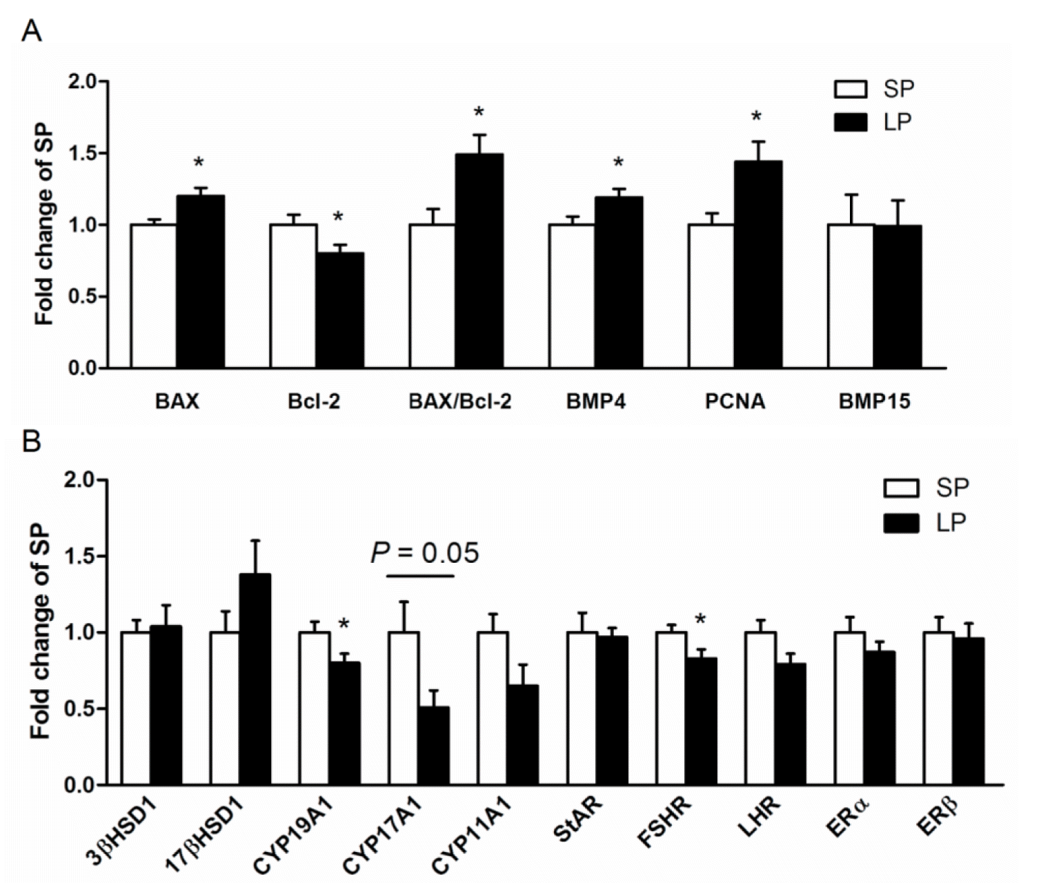
The LP piglets expressed significantly higher BAX (p<0.05) but lower Bcl-2 (p<0.05) mRNA in the ovary, which resulting in higher ratio of BAX/Bcl-2 (p<0.01). Moreover, mRNA expression of BMP4 (p<0.05) and PCNA (p<0.05) were significantly up-regulated in LP group (Figure 1A). Among seven steroidogenic genes detected, CYP17A1 (p = 0.05) tended to be lower, and CYP19A1 (p<0.05) were significantly down-regulated in LP piglets. Ovarian expression of ERα/β, 3βHSD, 17βHSD1, CYP11A1 and StAR mRNA in neonatal piglets was not affected by maternal dietary protein restriction. The alterations in ovarian expression of folliculogenic and steroidogenic genes were associated with significantly down-regulated FSHR mRNA expression (p<0.05) in the ovary of LP piglets (Figure 1B). These results suggest that maternal protein restriction affects the expression of folliculogenic and steroidogenic genes, which are related to follicle apoptosis and survival, in the ovary of neonatal offspring piglets.

Methylated DNA immunoprecipitation analysis in offspring neonatal piglets

To determine whether the alterations in ovarian gene expression involve modifications in CpG methylation on respective gene promoters, we employed MeDIP assay using specific primers to amplify the fragments spanning most of the CpG islands on gene promoters (Figure 2). However, no differences were detected in the methylation levels on BMP4, PCNA, or CYP19A1 gene promoters between SP and LP neonatal piglets ovary.

Expression of miRNAs predicted to target CYP19A1, FSHR, BMP4, and PCNA

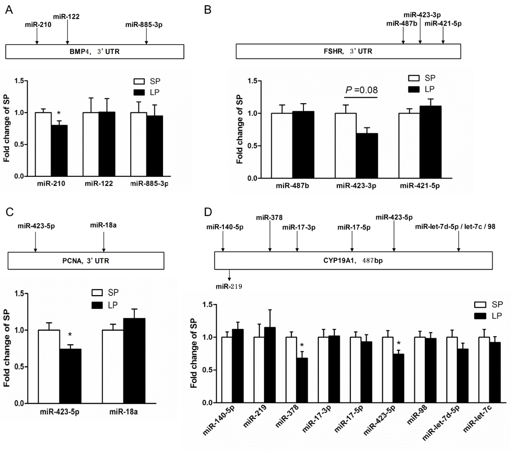
Nine miRNAs for CYP19A1, three for BMP4, two for PCNA and three for FSHR were chosen for miRNA quantification. We found that the expression of miR-423-5p targeting CYP19A1 and PCNA, miR-378 targeting CYP19A1 and miR-210 targeting BMP4 were significantly down-regulated (p<0.05) in the ovary of LP offspring. Moreover, the expression of miR-423-3ptargeting FSHR tended to be lower (p = 0.08) in the ovary of LP piglets (Figure 3). These results implicate that miRNAs-mediated post-transcriptional gene regulation is involved in the effects of maternal protein restriction on ovarian expression of folliculogenic and steroidogenic genes in the neonatal piglets.

DISCUSSION

To our knowledge, this is the first report on the effect of maternal LP diet on ovarian genes, miRNAs expression and MeDIP analysis in neonatal piglets. Alterations in the ovarian expression of folliculogenic and steroidogenic genes suggest the potential impact of maternal LP diet on ovarian development and function in neonatal piglets. Moreover, we provide preliminary evidences that miRNAs predicted to target folliculogenic and steroidogenic genes are involved in mediating the effect of maternal LP diet on ovarian function in newborn piglets.
Numerous studies in rats indicate that maternal malnutrition during pregnancy decreases ovarian weight in adults (da Silva Faria et al., 2004; Iwasa et al., 2011). In this study, disrupted ovarian development in prenatally undernourished offspring was observed in neonatal age; LP piglets demonstrated significantly lower ovary weight relative to body weight at birth. Sex steroid hormones are crucial for the development of ovary. In the present study, we examined the levels of sex steroid hormones and found that serum E2 concentration was higher in LP group. Other studies also reported significantly increased serum E2 concentration in 40-day-old rats subjected to maternal protein restriction during lactation (da Silva Faria et al., 2008; Faria et al., 2010). Primary follicles are the main sites for estrogen production in the neonatal animals, so higher E2 concentration may implicate increased number of primary follicles in the ovary of neonatal LP piglets (Faria et al., 2010).
The PCNA, a proliferation factor, is recently found to play an important role in ovarian folliculogenesis (Xu et al., 2011). In pigs, PCNA is expressed in growing follicles, but not in quiescent primordial follicles (Tománek and Chronowska, 2006). In the ovary of neonatal piglets, follicle development is mainly the transition from primordial to primary follicle (Ding et al., 2010). In this study, PCNA mRNA abundance was significantly higher in LP group than the SP group, suggesting that the ovary in neonatal LP piglets contains more primary follicles. This finding agrees with early reports that knock-down of PCNA expression by RNA interference retarded the transition of primordial follicles into primary follicles in vitro (Xu et al., 2011). The BMP4 can also promote the transition of primordial-to-primary follicles. Neonatal rat ovaries treated with exogenous BMP4 had a higher proportion of primary follicles and fewer primordial follicles (Nilsson and Skinner, 2003). Conversely, immune-neutralization with BMP-4 antibody led to lower primary follicles and higher primordial follicles in mouse ovaries (Tanwar et al., 2008). Here, we found that the change of BMP4 expression was consistent with PCNA. These findings indicate that the ovary development in neonatal LP piglets may be accelerated. It also may help to explain the observations that maternal undernutrition in pregnancy results in premature aging of offspring ovary (Bernal et al., 2010) and an earlier termination of breeding capacity (da Silva Faria et al., 2008).
Apoptosis is a physiological process which is required for primordial follicle assembly and early follicle development (Ding et al., 2010). The PCNA can also regulate primordial follicle assembly in neonatal mouse ovaries by promoting apoptosis of oocytes (Xu et al., 2011). In mammals, follicle apoptosis or follicular atresia begins from early fetal period with most follicles lost at birth (Ding et al., 2010). These reports suggest that excessive ovarian follicular development in neonatal piglets accompanied with increased apoptosis of follicle. The ratio of proapoptotic factor BAX and antiapoptotic factor Bcl-2 is an evaluation index for the level of apoptosis (Rucker et al., 2000; Sun et al., 2012). In this study, the ratio of BAX/Bcl-2 was higher in the ovary of LP piglets. The enhanced follicle apoptosis further supporting our presumption that maternal LP diet advances the early follicle development in the ovary of neonatal pigs.
Estrogen has multiple effects on the development of ovarian follicles through its receptors (ERα/β). Studies on rat ovaries indicate that E2 inhibits primordial to primary follicle transition both in vivo and in vitro (Kezele and Skinner, 2003). In this study, ovarian expression of ERα/β mRNA was not altered, despite significantly elevated serum E2 concentration in LP piglets. The elevated serum E2 level may result from accelerated ovary development in LP piglets. Therefore, enhanced steroidogenesis may be expected in the ovary of LP piglets. On the contrary, the ovarian mRNA expression of estrogen biosynthesis genes, such as CYP17A1 and CYP19A1, tended to be or are significantly down-regulated in LP piglets, which was associated with a significant down-regulation of FSHR mRNA. This contradicts with the elevated serum E2 and advanced ovarian follicular development in LP piglets. In addition, there is no difference in the DNA methylation of PCNA, BMP4, and CYP19A1 promoter. So it is possible that the protein content or the enzyme activity is uncoupled with the mRNA abundance, with the possible involvement of post-transcriptional regulation. Unfortunately, we were not able to detect the protein content or the enzyme activity because neonatal ovaries are not big enough for both RNA and protein extractions.
However, we detected the miRNA regulation, and found that the expression of miR-423-5p and miR-378 targeting CYP19A1 was significantly downregulated, implying that the post-transcriptional repression of CYP19A1 may be decreased which thereby results in higher CYP19A1 protein content or enzyme activity. Similar alterations are also detected for miRNAs targeting folliculogenic genes. MiR-423-5p and miR-210 which are predicted to target PCNA and BMP4 respectively were also found to be significantly down-regulated in the ovary of LP piglets, suggesting an inhibitory role of miRNAs in mRNA stability. It is noted that miR-423-5p is predicted to target both CYP19A1 and PCNA, but the regulatory mechanisms may be different, being translation inhibition for CYP19A1 while mRNA degradation for PCNA. We did not detect alterations in the expression of the two miRNAs predicted to target porcine FSHR gene, but whether other miRNAs are involved in the regulation of FSHR remains elusive. There is no 3′ UTR sequence reported for porcine CYP17A1, BAX, and Bcl-2 genes, therefore we are not able to predict their regulatory miRNAs from 3′ UTR sequences.
In conclusion, our results suggest that a maternal LP diet in pregnancy affects the expression of folliculogenic and steroidogenic genes in neonatal piglets. Post-transcriptional regulation of gene expression by miRNAs plays an important role in these processes. Neonatal changes in ovarian function may persist to adulthood, with long-term consequences on folliculogensis and steroidogensis. Our findings may shed light on understanding the ovarian aging and ovarian reserve reduction in prenatally undernourished offspring.

ACKNOWLEDGMENTS

This work was supported by the National Basic Research Program of China (2012CB124703, 2014CB138502), the Special Fund for Agro-scientific Research in the Public Interest (201003011), the Fundamental Research Funds for the Central Universities (KYZ200913) and the Priority Academic Program Development of Jiangsu Higher Education Institutions.

Figure 1
Ovarian expression of folliculogenic and steroidogenic genes in neonatal piglets. A, folliculogenic genes expression; B, steroidogenic genes expression. Data are expressed as mean±standard error of the mean. Asterisks indicate statistically significant differences (p<0.05), n = 8.
ajas-27-12-1695f1.gif
Figure 2
DNA methylation analysis in the CpG islands of BMP4, PCNA, and CYP19A1 promoter. The CpG islands in the promoter of each gene are presented above the bargraphs of respective MeDIP analysis. A, CpG islands of BMP4 promoter and MeDIP analysis; B, CpG island of PCNA promoter and MeDIP analysis; C, CpG island of CYP19A1 promoter and MeDIP analysis. Data are expressed as mean±standard error of the mean. Asterisks indicate statistically significant differences (p<0.05), n = 8. CpG, cytosine phosphate guanine; BMP4, bone morphogenic protein 4; PCNA, proliferating cell nuclear antigen; CYP19A1, cytochrome P450 aromatase; MeDIP, methylated DNA immunoprecipitation.
ajas-27-12-1695f2.gif
Figure 3
Ovarian expression of miRNAs predicted to target A, BMP4; B, FSHR; C, PCNA; and D, CYP19A1 genes in neonatal piglets. Predicted binding sites of miRNAs on each target gene are presented above the bargraphs of respective miRNA expression. Data are expressed as mean±standard error of the mean. Asterisks indicate statistically significant differences (p<0.05), n = 8. BMP4, bone morphogenic protein 4; FSHR, follicle-stimulating hormone receptor; PCNA, proliferating cell nuclear antigen; CYP19A1, cytochrome P450 aromatase.
ajas-27-12-1695f3.gif
Table 1
Ingredients and calculated composition of the experimental diets
Items Standard protein Low-protein
Ingredient (g/kg)
 Corn 370 615
 Wheat 300 100
 Bran 80 100
 Soybean Meal 170 0
 Maize starch 0 100
 Lignocellulose 30 40
 CaHPO4 20 25
 Soybean oil 10 0
 Premix* 20 20
Calculated composition
 Digestible energy (MJ/kg) 13.10 13.10
 Crude protein (%) 15 7.50
 Crude fiber (%) 4.50 4.30
 Calcium (%) 0.84 0.85
 Phosphorous (%) 0.65 0.61

* The premix contains (per kilogram): retinol: 1,100 kIU; cholecalciferol, 350 kIU; vitamin K3, 0.4 g; vitamin B1, 0.4 g; vitamin B2, 1,640 mg; vitamin B6, 0.65 g; vitamin B12, 4.4 mg; lysine, 72 g; niacin, 4.5 g; pantothenic acid, 2.5 g; d-pantothenic acid, 2 g; folic acid, 5.2 g; biotin, 30 mg; d-biotin, 16 mg; choline chloride, 30 g; vitamin C, 20 g; manganese, 0.8 g; zinc, 7 g; ferrous, 7 g; copper, 2 g; selenium, 20 mg; sodium chloride, 3 g; β-xylanase, 8,000 kIU; antioxidant, 0.19 g; acidifier, 2.5 g.

Table 2
Nucleotide sequences of specific primers
Gene name Products (bp) Primer sequences (5′ to 3′) GenBank accession number
18S rRNA 122 F: CCCACGGAATCGAGAAAGAG
R: TTGACGGAAGGGCACCA
AY265350.1
BAX 143 F: TGACGGCAACTTCAACTGGG
R: GCAGCCGATCTCGAAGGAA
XM_003127290.2
Bcl-2 142 F: GCCTTTGTGGAGCTGTATGG
R: CCCGTGGACTTCACTTATGG
XM_003121700.2
BMP15 235 F: TCAGAGCCACTGTGGTTTATCG
R: CACATGAAGCGGAGTCGTAGAA
NM_001005155.1
BMP4 172 F: GGGCTCGGAAGAAGAATAAGA
R: ACGATGGCATGATTGGTTGA
NM_001101031.2
PCNA 90 F: GGTTCAGGCTCTGTTGTGGTGT
R: GCTACAGGTGGAAGAAAGAAGGAA
XM_003359883.1
3βHSD1 145 F: CACTGACCTGGGCTGATGAC
R: GTGGCGAGAAGCAGACAAGA
NM_001004049.1
17βHSD2 196 F: TCGAGCAGACGTTCCTTGAG
R: TGGTCGCCGAACACTCTTT
NM_001128472.1
CYP19A1 187 F: GCTGCTCATTGGCTTAC
R: TCCACCTATCCAGACCC
NM_214431.1
CYP17A1 136 F: TCCAAGCCAAGACGAAC
R: TTTACCACAGAGGCAGAAG
NM_214428.1
CYP11A1 149 F: GGCTCCAGAGGCCATAAAGA
R: ACTCAAAGGCGAAGCGAAAC
X13768.1
StAR 202 F: GACTTTGTGAGTGTCGGCTGTA
R: ATCCCTTGAGGTCAATGCTG
U53020.1
FSHR 152 F: TCACAGTCCCTCGGTTCCTT
R: AGCATCACAGCCTGCTCCA
NM_214386.2
LHR 255 F: ATGGGGCTCTACCTGCTACTCA
R: GAGCCACCCTCCAAGCATAA
NM_214449.1
ERα 151 F: ATGAAGTGCAAGAACGTGGTG
R: AATGCGATGGAGTTGAGCC
NM_214220.1
ERβ 191 F: GCCCTGTTACCAGTCCAAGTT
R: ACGCCGGTTCTTATCTATCGT
NM_001001533.1

F, forward; R, reverse; BAX, bcl-2 associated X protein; Bcl-2, B cell lymphoma/leukemia-2; BMP, bone morphogenic protein; PCNA, proliferating cell nuclear antigen; βHSD, β-hydroxysteroid dehydrogenase; CYP19A1, cytochrome P450 aromatase; CYP17A1, cytochrome P450 17a-hydroxylase; CYP11A1, cytochrome P450 cholesterol side-chain cleavage enzyme; StAR, steroidogenic acute regulatory protein; FSHR, follicle-stimulating hormone receptor; LHR, luteinizing hormone receptor; ER, estrogen receptor α.

Table 3
The primer sequences of miRNAs expression
Name Primer sequences (5′ to 3′) miRbase accession
miR-378 ACTGGACTTGGAGTCAGAAGGC MIMAT0013868
miR-98 TGAGGTAGTAAGTTGTATTGTT MIMAT0013905
miR -let-7d-5p AGAGGTAGTAGGTTGCATAGTT MIMAT0025356
miR-140-5p AGTGGTTTTACCCTATGGTAG MIMAT0002143
miR-140-3p TACCACAGGGTAGAACCACGGAC MIMAT0013881
miR-let-7c TGAGGTAGTAGGTTGTATGGTT MIMAT0002151
miR-423-5p TGAGGGGCAGAGAGCGAGACTTT MIMAT0013880
miR-423-3p AGCTCGGTCTGAGGCCCCTCAGT MIMAT0013881
miR-17-5p CAAAGTGCTTACAGTGCAGGTAG MIMAT0007755
miR-17-3p ACTGCAGTGAAGGCACTTGTAG MIMAT0015268
miR-885-3p AGGCAGCGGGGTGTAGTGGAT MIMAT0013903
miR-421-5p CCTCATTAAATGTTTGTTGAATGA MIMAT0017970
miR-18a TAAGGTGCATCTAGTGCAGATA MIMAT0002161
miR-122 TGGAGTGTGACAATGGTGTTTGT MIMAT0002119
miR-210 CTGTGCGTGTGACAGCGGCTGA MIMAT0007761
miR-487b GTGGTTATCCCTGTCCTGTTCG MIMAT0017973
U6 snRNA GGCAAGGATGACACGCAAAT ENSSSCT00000019750
poly(T) adapter TAGAGTGAGTGTAGCGAGCACAGAATTAATACGACTCACTATAGGTTTTTTTTTTTTTTTTVN
universal primer TAGAGTGAGTGTAGCGAGCA
Table 4
The primer sequences of MeDIP analysis
Name Primer sequences (5′ to 3′)
BMP4 CpG1 F: AACTGGGAACAGGCTTAGGAA
R: GACCCTCGAAACTGAATCTGC
BMP4 CpG2 F: GACCTGCCTTCCAGAGTCCA
R: CCGCAGTCAAGTGGGAGAA
PCNA CpG F: CCGCATCTGCAACCTATACCA
R: CGAATCCGACTAGGAACCATG
CYP19A1 CpG F: ATCGTGGAGCAGTGGTTA
R: GTCGAATCGGAGCCTTAG
mc1 F: CCCTATAACGCCTTGCCAAACC
R: GGGTAGGTGCCTGCTTTCGTAG

MeDIP, methylated DNA immunoprecipitation; F, forward; R, reverse; BMP, bone morphogenic protein; PCNA, proliferating cell nuclear antigen; CYP19A1, cytochrome P450 aromatase; mc1, methylation control 1.

Table 5
Body weight, ovary weight and serum concentrations of sex steroid hormones in neonatal piglets
Parameter SP
n = 8
LP
n = 8
p value
BW (kg) 1.49±0.08 1.57±0.04 0.40
OW (g) 0.11±0.01 0.08±0.01 0.07
OW/BW (g/kg) 0.08±0.01 0.05±0.01 0.03
E2 (pg/mL) 6.42±2.12 38.33±17.57 0.04
P4 (ng/mL) 9.10±1.42 13.56±2.94 0.16
T (ng/mL) 0.42±0.38 1.16±0.68 0.33

SP, standard protein; LP, low-protein; BW, body weight; OW, ovary weight; E2, 17β-estradiol; P4, progesterone; T, testosterone; SEM, standard error of the mean.

Values are mean±SEM, statistical significances were set at p<0.05.

REFERENCES

Baley J, Li J. 2012. Micrornas and ovarian function. J Ovarian Res 5:8
crossref pmid pmc
Bernal AB, Vickers MH, Hampton MB, Poynton RA, Sloboda DM. 2010. Maternal undernutrition significantly impacts ovarian follicle number and increases ovarian oxidative stress in adult rat offspring. Plos One 5:12e15558
crossref pmid pmc
Camp TA, Rahal JO, Mayo KE. 1991. Cellular localization and hormonal regulation of follicle-stimulating hormone and luteinizing hormone receptor messenger rnas in the rat ovary. Mol Endocrinol 5:1405–1417.
crossref pmid
Chen Y, Jefferson WN, Newbold RR, Padilla-Banks E, Pepling ME. 2007. Estradiol, progesterone, and genistein inhibit oocyte nest breakdown and primordial follicle assembly in the neonatal mouse ovary in vitro and in vivo. Endocrinology 148:3580–3590.
crossref pmid
da Silva Faria T, da Fonte Ramos C, Sampaio FJB. 2004. Puberty onset in the female offspring of rats submitted to protein or energy restricted diet during lactation. J Nutr Biochem 15:123–127.
crossref pmid
da Silva Faria T, de Bittencourt Brasil F, Sampaio FJ, da Fonte Ramos C. 2008. Maternal malnutrition during lactation alters the folliculogenesis and gonadotropins and estrogen isoforms ovarian receptors in the offspring at puberty. J Endocrinol 198:625–634.
crossref pmid
Ding W, Wang W, Zhou B, Zhang W, Huang P, Shi F, Taya K. 2010. Formation of primordial follicles and immunolocalization of PTEN, PKB and FOXO3a proteins in the ovaries of fetal and neonatal pigs. J Reprod Dev 56:162–168.
crossref pmid
Durlej M, Knapczyk-Stwora K, Duda M, Galas J, Slomczynska M. 2011. The expression of FSH receptor (FSHR) in the neonatal porcine ovary and its regulation by flutamide. Reprod Domest Anim 46:377–384.
crossref pmid
Faria TS, Brasil FB, Sampaio FJB, Ramos CF. 2010. Effects of maternal undernutrition during lactation on estrogen and androgen receptor expressions in rat ovary at puberty. Nutrition 26:993–999.
crossref pmid
Fiedler SD, Carletti MZ, Hong X, Christenson LK. 2008. Hormonal regulation of microrna expression in periovulatory mouse mural granulosa cells. Biol Reprod 79:1030–1037.
crossref pmid pmc
Fire A, Xu S, Montgomery MK, Kostas SA, Driver SE, Mello CC. 1998. Potent and specific genetic interference by double-stranded rna in caenorhabditis elegans. Nature 391:6669806–811.
crossref pmid
Fortune J, Cushman R, Wahl C, Kito S. 2000. The primordial to primary follicle transition. Mol Cell Endocrinol 163:53–60.
crossref pmid
Godfrey KM, Gluckman PD, Hanson MA. 2010. Developmental origins of metabolic disease: Life course and intergenerational perspectives. Trends Endocrin Metab 21:199–205.
crossref
Grzesiak M, Knapczyk-Stwora K, Duda M, Slomczynska M. 2012. Elevated level of 17β-estradiol is associated with overexpression of FSHR, CYP19A1, and CTNNB1 genes in porcine ovarian follicles after prenatal and neonatal flutamide exposure. Theriogenology 78:2050–2060.
crossref pmid
Ibáñez L, Potau N, Ferrer A, Rodriguez-Hierro F, Marcos MV, de Zegher F. 2002. Reduced ovulation rate in adolescent girls born small for gestational age. J Clin Endocrinol Metab 87:3391–3393.
crossref pmid
Iwasa T, Matsuzaki T, Murakami M, Kinouchi R, Gereltsetseg G, Yamamoto S, Kuwahara A, Yasui T, Irahara M. 2011. Delayed puberty in prenatally glucocorticoid administered female rats occurs independently of the hypothalamic Kiss1-Kiss1R-GnRH system. Int J Dev Neurosci 29:183–188.
crossref pmid
Jia Y, Cong R, Li R, Yang X, Sun Q, Parvizi N, Zhao R. 2012. Maternal low-protein diet induces gender-dependent changes in epigenetic regulation of the glucose-6-phosphatase gene in newborn piglet liver. J Nutr 142:1659–1665.
crossref pmid
Kertesz M, Iovino N, Unnerstall U, Gaul U, Segal E. 2007. The role of site accessibility in microrna target recognition. Nat Genet 39:1278–1284.
crossref pmid
Kezele P, Skinner MK. 2003. Regulation of ovarian primordial follicle assembly and development by estrogen and progesterone: Endocrine model of follicle assembly. Endocrinology 144:3329–3337.
crossref pmid
Liu X, Wang J, Li R, Yang X, Sun Q, Albrecht E, Zhao R. 2011. Maternal dietary protein affects transcriptional regulation of myostatin gene distinctively at weaning and finishing stages in skeletal muscle of Meishan pigs. Epigenetics 6:899–907.
crossref pmid
Meikle D, Westberg M. 2001. Maternal nutrition and reproduction of daughters in wild house mice (Mus musculus). Reproduction 122:437–442.
crossref pmid
Nilsson EE, Skinner MK. 2003. Bone morphogenetic protein-4 acts as an ovarian follicle survival factor and promotes primordial follicle development. Biol Reprod 69:1265–1272.
crossref pmid
Rae MT, Kyle CE, Miller DW, Hammond AJ, Brooks AN, Rhind SM. 2002. The effects of undernutrition, in utero, on reproductive function in adult male and female sheep. Anim Reprod Sci 72:63–71.
crossref pmid
Rucker EB, Dierisseau P, Wagner KU, Garrett L, Wynshaw-Boris A, Flaws JA, Hennighausen L. 2000. Bcl-x and bax regulate mouse primordial germ cell survival and apoptosis during embryogenesis. Mol Endocrinol 14:1038–1052.
crossref pmid
Sirotkin AV, Ovcharenko D, Grossmann R, Lauková M, Mlynček M. 2009. Identification of micrornas controlling human ovarian cell steroidogenesis via a genome-scale screen. J Cell Physiol 219:415–420.
crossref pmid
Sloboda DM, Hickey M, Hart R. 2011. Reproduction in females: The role of the early life environment. Hum Reprod Update 17:210–227.
crossref pmid
Sloboda DM, Howie GJ, Pleasants A, Gluckman PD, Vickers MH. 2009. Pre- and postnatal nutritional histories influence reproductive maturation and ovarian function in the rat. Plos One 4:8e6744
crossref pmid pmc
Sun R, Lei L, Cheng L, Jin ZF, Zu SJ, Shan ZY, Wang ZD, Zhang JX, Liu ZH. 2010. Expression of GDF-9, BMP-15 and their receptors in mammalian ovary follicles. J Mol Histol 41:325–332.
crossref pmid
Sun Y, Lin Y, Li H, Liu J, Sheng X, Zhang W. 2012. 2, 5-hexanedione induces human ovarian granulosa cell apoptosis through bcl-2, bax, and caspase-3 signaling pathways. Arch Toxicol 86:205–215.
crossref pmid
Tanwar PS, O’Shea T, McFarlane JR. 2008. In vivo evidence of role of bone morphogenetic protein-4 in the mouse ovary. Anim Reprod Sci 106:232–240.
crossref pmid
Tománek M, Chronowska E. 2006. Immunohistochemical localization of proliferating cell nuclear antigen (PCNA) in the pig ovary. Folia Histochem Cytobiol 44:269–274.
pmid
Torley KJ, da Silveira JC, Smith P, Anthony RV, Veeramachaneni D, Winger QA, Bouma GJ. 2011. Expression of mirnas in ovine fetal gonads: Potential role in gonadal differentiation. Reprod Biol Endocrinol 9:2
crossref pmid pmc
Xu B, Hua J, Zhang Y, Jiang X, Zhang H, Ma T, Zheng W, Sun R, Shen W, Sha J, Cooke HJ, Shi Q. 2011. Proliferating cell nuclear antigen (PCNA) regulates primordial follicle assembly by promoting apoptosis of oocytes in fetal and neonatal mouse ovaries. Plos One 6:1e16046
crossref pmid pmc
TOOLS
METRICS Graph View
  • 23 Crossref
  • 26 Scopus
  • 11,751 View
  • 118 Download
Related articles


Editorial Office
Asian-Australasian Association of Animal Production Societies(AAAP)
Room 708 Sammo Sporex, 23, Sillim-ro 59-gil, Gwanak-gu, Seoul 08776, Korea   
TEL : +82-2-888-6558    FAX : +82-2-888-6559   
E-mail : editor@animbiosci.org               

Copyright © 2024 by Asian-Australasian Association of Animal Production Societies.

Developed in M2PI

Close layer
prev next