Go to Top Go to Bottom
Lim, Seo, Choi, Yoon, Nam, Kim, Cho, and Chang: Metagenome Analysis of Protein Domain Collocation within Cellulase Genes of Goat Rumen Microbes

Abstract

In this study, protein domains with cellulase activity in goat rumen microbes were investigated using metagenomic and bioinformatic analyses. After the complete genome of goat rumen microbes was obtained using a shotgun sequencing method, 217,892,109 pair reads were filtered, including only those with 70% identity, 100-bp matches, and thresholds below E−10 using METAIDBA. These filtered contigs were assembled and annotated using blastN against the NCBI nucleotide database. As a result, a microbial community structure with 1431 species was analyzed, among which Prevotella ruminicola 23 bacteria and Butyrivibrio proteoclasticus B316 were the dominant groups. In parallel, 201 sequences related with cellulase activities (EC.3.2.1.4) were obtained through blast searches using the enzyme.dat file provided by the NCBI database. After translating the nucleotide sequence into a protein sequence using Interproscan, 28 protein domains with cellulase activity were identified using the HMMER package with threshold E values below 10−5. Cellulase activity protein domain profiling showed that the major protein domains such as lipase GDSL, cellulase, and Glyco hydro 10 were present in bacterial species with strong cellulase activities. Furthermore, correlation plots clearly displayed the strong positive correlation between some protein domain groups, which was indicative of microbial adaption in the goat rumen based on feeding habits. This is the first metagenomic analysis of cellulase activity protein domains using bioinformatics from the goat rumen.

INTRODUCTION

Goats have an extremely varied diet including the tips of woody shrubs, trees, and lignocellulosic agricultural byproducts. Symbiont microbes in the rumen of these herbivores play key roles in providing the hosts with various nutrients. Enzymes secreted by rumen microbes are essential for the conversion of cellulose and hemi-cellulose into simple sugars, which are metabolized to volatile fatty acids by rumen microbes. Produced volatile fatty acids serve as energy sources for ruminants. Many studies have investigated the symbiotic microorganisms in the rumen because of their link to economically or environmentally important traits such as feed conversion efficiency, methane production (Hegarty, 1999; Guan et al., 2008; Hess et al., 2011). There have been various studies about the correlation between rumen microbiota and their role in nutrients digestion for sheep and cattle. Especially, information for the microbial digestion consortia in goat rumen was expected to provide its species distinct characteristics compared to those of other ruminant animals (McAllister et al., 1994).
A key challenge in this study was identifying rumen microbial profiles, which are associated and potentially predictive of these traits. Thus, methods for profiling the rumen microbial population should be relatively inexpensive and efficient to allow a large number of individuals to be profiled (Ross et al., 2012). Untargeted rumen bacterial communities contain numerous novel gene sequences based on deep sequencing of pooled samples of true biological variation. The rumen metagenome profile included the counts of reads that aligned to each contig, which could be analyzed using metagenomic tools and correlation plots. The composition of the microbial population differs between goat species and based on their diet. Analysis of microorganisms in the rumen fluid of different herbivores revealed bacteria (1010 to 1011 cells/ml, representing more than 50 genera), ciliate protozoa (104 to 106/ml, from 25 genera), anaerobic fungi (103 to 105 zoospores/ml, representing six genera), and bacteriophages (108 to 109 ml). These numbers represented only a small fraction of the microbial species in rumens of animals on fiber-based diets since less than 10 to 20% of microbial populations are cultivable on synthetic media (Zhou et al., 2011). However, metagenomic research has generated genetic information on the entire microbial community, which is important because 99% of microbes cannot be isolated or cultured. The metagenomic method provides a global microbial gene pool without the need to culture of the microorganisms. In this study, we analyzed the complete genome of goat rumen microbes obtained using a shotgun sequencing method. This differed from previous studies on microbes based on 16 rRNA. Also, our results were filtered under strict conditions and provided high-quality results on the rumen microbe community and cellulase activity protein domains.

MATERIALS AND METHODS

Sampling and extraction of genomic DNA

Rumen fluid was collected from a 1-yr-old Korean native goat and Saanen hybrid raised on Timothy (Phleumpratense) hay at a private goat farm in the Cheonan City area and slaughtered at a local slaughter house. Rumen fluid was filtered through four layers of cheesecloth. Genomic DNA was isolated from rumen fluid using the Wizard Genomic DNA Purification Kit (Promega, US) according manufacturer’s protocol. Gel electrophoresis was performed with 1% agarose gel at 50 V for 2 h to check both quality and quantity of isolated genomic DNA.

DNA shotgun paired-end library preparation

Random DNA fragmentation was performed using the Covaris S2 System, and the DNA library was prepared using TruSeqDNA Sample Prep. Kit (Illumina, US). Briefly, DNA fragments were repaired to blunt-ended DNA by fill-in and exonuclease after A-tailing was conducted to prevent the formation of adapters, dimers, and concatemers. Adaptors were ligated to genomic DNA inserts at a molar ratio of 10:1. The DNA samples were then amplified via polymerase chain reaction (PCR) using two universal primers. One primer contained an attachment site for the flow cell and the other contained sequencing sites for the index read. After gel electrophoresis of the PCR product, 600 to 700-bp fragments (including the insert and adapter) were selected and purified for genomic sequencing.

Genomic sequencing

Genomic DNA sequences were generated using the Illumina Hiseq2000 platform. Briefly, only library fragments with proper adapters at both ends were amplified using P5 and P7 primers on the flow cell. Clonal clusters were generated using TreSeq PE Cluster kitV3-cBot-HS (ILPE-401-3001; Illumina). Using the HiSeq2000 platform with TruSeq SBS Kit v3-HS (200 cycles; ILFC-401-3001; Illumina) 435,784, 218 reads were obtained.

Metagenomic bioinformatics application

Each pair read, scaffold, and contig of the shotgun sequencing of goat rumen microbes was summarized in Supplementary Figure 1 and Supplementary Table 1. Whole genomic DNA of collected goat rumen microbes were extracted for Illumina sequencing without DNA targeting. This shotgun sequencing generated 217,892,109 pair reads, which were filtered based on 70% identity, more than a 100-bp match, and a threshold below E−10 based on METAIDBA (Peng et al., 2011). These filtered 1,373,011 scaffolds were assembled and annotated to 114,031 contigs using blastN against the NCBI nucleotide database. The domains of these 201 protein sequences were assigned to the cellulase (EC.3.2.1.4) database and translated using the HMMER package with threshold E values below 10−5. Finally, these annotated genomic sequences were assigned for both identification of microbial species and cellulase-like protein domains

RESULTS AND DISCUSSION

Microbial community structure in goat rumen

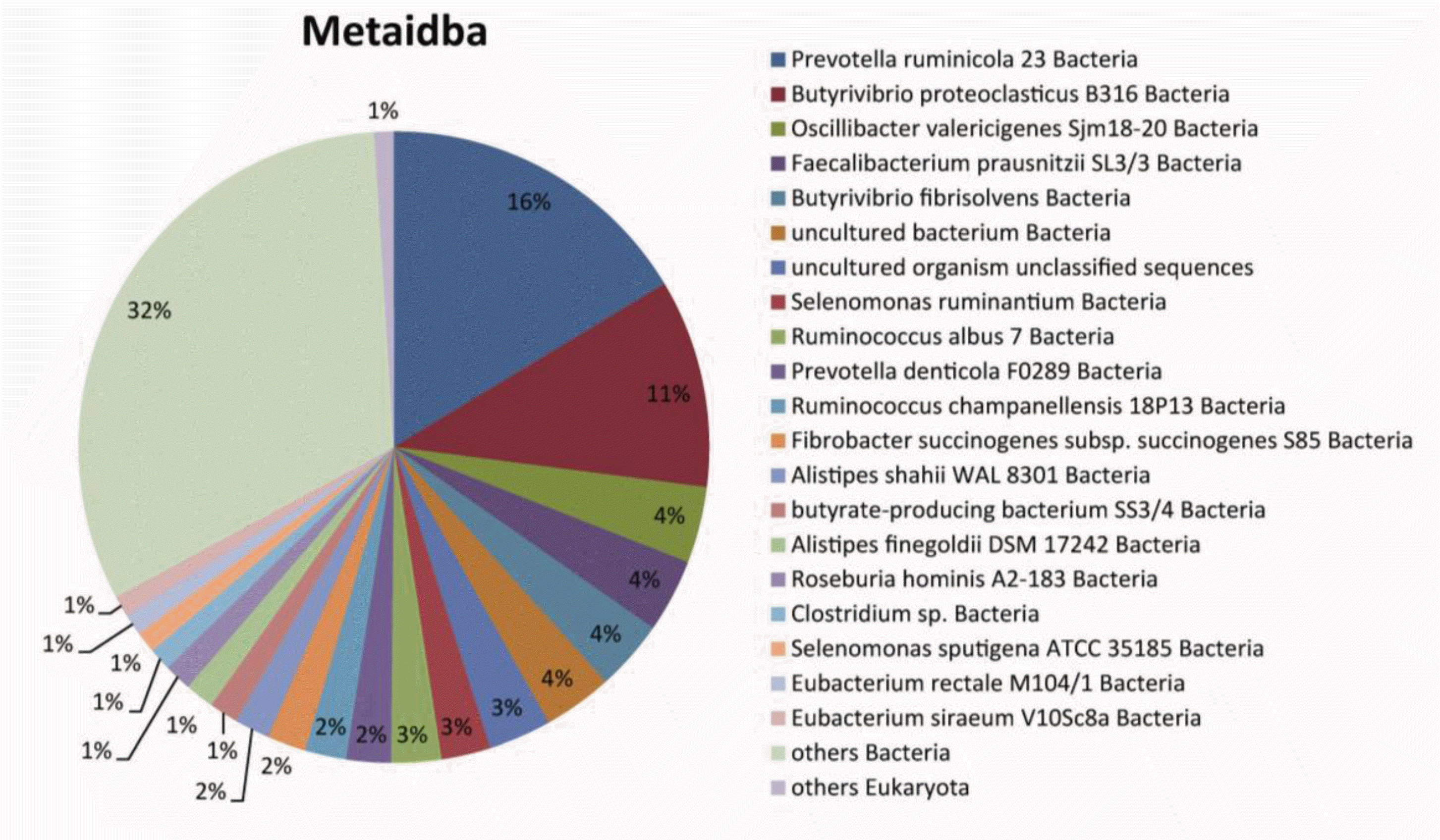
The isolated genes in rumen fluid were classified into a total of 1,704 organisms, among which each 181 and 1431 ID corresponded to plant and bacteria, respectively. Using the METAIDBA metagenomic bioinformatic program, 114,031 sequences were classified into 1431 species; their population structure at the species level is graphically depicted in Figure 1. Prevotella ruminicola 23 bacteria and Butyrivibrio proteoclasticus B316 bacteria were the dominant populations, accounting for 16% and 11%, respectively.
The majority of goat rumen bacteria identified in this study have been previously reported in the rumens of cow or lamb, such as Prevotella ruminicola 23, Butyrivibrio proteoclasticus B316, and Butyrivibrio fibrisolvens (Bryant and Small, 1956; Van Gylswyk and Van Der Toorn, 1986; McKain et al., 1992; Moon et al., 2008). Also, some microorganisms such as butyrate-producing bacterium SS3/4 have been identified in the human colon. Previous studies have revealed the detailed rumen metabolism of Fibrobacter succinogenes subsp. Fibrobacter succinogenes S85 and Selenomonas ruminantium (Heinrichova et al., 1989; Chow and Russell, 1992).

Protein domains with cellulase activity

Cellulase protein ID was obtained from the enzyme.dat file provided by the NCBI database. As a result, 201 sequences related with cellulase activity were obtained through blast searches using the NCBI BLAST program. In total, 28 protein domains with cellulase activity are summarized in Table 1. For other ruminant animals, Toyoda and coworkers analyzed the cellulose-binding proteins from sheep rumens, which consisted of endo-glucanases, proteins from fiber degrading bacterium and exo-glucanases, respectively (Toyoda et al., 2009). For cattle, constructed metagenomic library and identified 22 clones with distinct hydroylic activities such as 12 esterases, nine endo-β-1,4-glucanases and one cyclodextrin (Ferrer et al., 2005). Considering the close correlation between rumen microbial ecology and its enzymatic functions according to the other ruminal livestock (Krause et al., 2013), list of cellulase-like protein domain list of this study can provide a clue to the characterization of Korean native goat rumen.

Profile of protein domains with cellulase activity

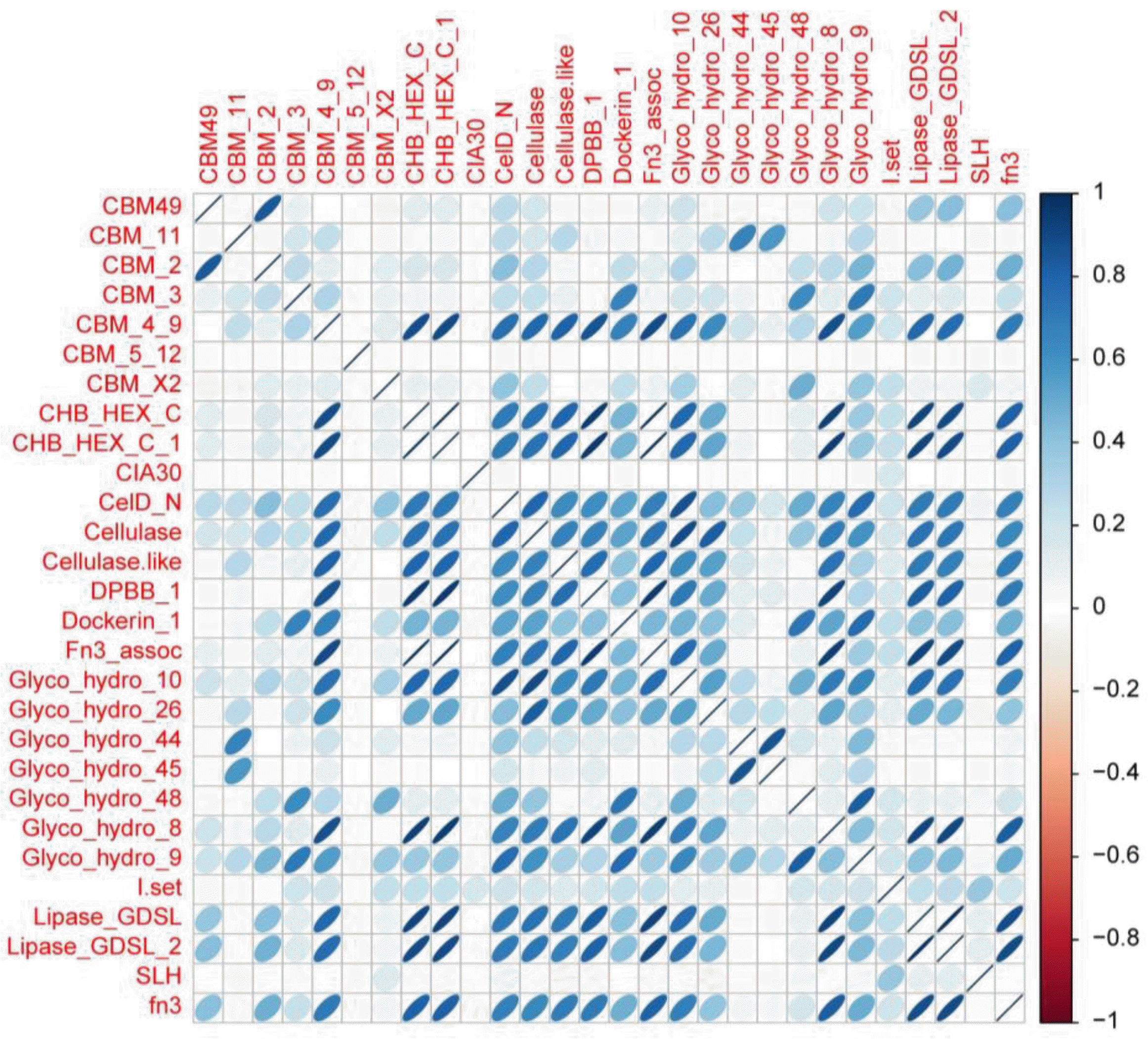
After 28 protein domains with cellulase activity were identified, the richness of each domain was analyzed (Supplementary Figure 2). Some of protein domains were overlapped to same part of sequences and also counted. The dominant bacteria had a larger number of protein domains, which suggested that strong cellulase activities were related to bacterial survival in the goat rumen. Protein domains with high richness such as lipase GDSL, cellulase, and Glyco hydro 10 were also identified in the goat rumen microbes. Both lipase GDSL and lipase GDSL_2 have been reported to have molecular function of cellulose binding (Galagan et al., 2005; Wortman et al., 2009). This is speculated as one of the main reason for its high detection. Next, the number of protein domains in each microbe was investigated (Supplementary Figure 3). Prevalent bacteria such as Prevotella ruminicola 23 bacteria and Butyrivibrio protein domain ratio in each bacterial species, of which proteoclasticus B316 contained a large number of cellulase definition was the portion of cellulase-like protein domain protein domains, implying that these bacteria play a role in to the assembled and annotated contigs, was analyzed to the degradation of cellulose in the goat rumen. Finally, the evaluate the richness of protein domain with cellulase activity in the dominant bacterial species (Supplementary Figure 4). The dominant bacteria showed a ratio greater than 1, suggesting that they have high cellulase activity. A correlation plot among 28 protein domains (Figure 2) confirmed the strong positive correlation between some protein domain groups. For example, CHB_HEX_c and CHB_HEX_c-1, CHB_HEX_c and fn3 asso, and CHB_HEX_c -1 and fn3 asso had a positive correlation greater than 0.99.
Another group of lipase GDSLs, lipase GDSL_2, also showed a positive correlation greater than 0.99. To determine whether the goat rumen microbe profile was predictive of the rumen fluid metagenome profile, we correlated every rumen metagenome profile with every cellulase activity protein domain. We then determined whether the correlations were higher for samples from the same animal than for between animal samples. The results suggested that rumen fluid samples had strong correlations with each protein domain. Microbial community structure and specific protein domains with cellulase activity in the goat rumen have been identified using metagenomic analysis with both shotgun sequencing and bioinformatics. This study demonstrated that specific dominant bacterial species and protein domains have strong positive correlations, suggesting adaption to the unique feeding habits of goats.

CONCLUSIONS

In this study, microbial community structure and specific protein domains with cellulase activity in the goat rumen were identified using metagenomic analysis with both shotgun sequencing and bioinformatics. As a result, the presence of both specific dominant bacterial species such as Prevotella ruminicola 23, Butyrivibrio proteoclasticus B316, and Butyrivibrio fibrisolvens were identified among 1,431 bacteria in rumen fluid. At the same time, 28 protein domains with cellulase-like activity such as lipase GDSL, cellulase, and Glyco hydro 10 were identified with strong positive correlations, suggesting adaption to the unique feeding habits of goats.

Supplementary Materials

Supplementary Figure 1
Each pair read, scaffold and contig from the shotgun sequencing of goat rumen microbes.
ajas-26-8-1144-11f3.tif
Supplementary Figure 2
Richness of each protein domain with cellulase activity in the goat rumen microbial population.
ajas-26-8-1144-11f4.tif
Supplementary Figure 3
Protein domains with cellulase activity were present in over 1% of the dominant microbe species.
ajas-26-8-1144-11f5.tif
Supplementary Figure 4
Ratio of protein domain in each bacteria species.
ajas-26-8-1144-11f6.tif
Supplementary Table 1
Assembly and annotation statistics
Assembly Annotation
Total pair reads 217,892,109
Scaffolds 1,373,011
Contigs 114,031

ACKNOWLEDGMENTS

This work was supported by a grant PJ0081912013 from Bio-Green 21 Program, Rural Development Administration, Republic of Korea.

Figure 1
Analysis of the goat rumen microbial community structure at the species level.
ajas-26-8-1144-11f1.gif
Figure 2
Correlation plot between each cellulase activity protein domain in goat rumen.
ajas-26-8-1144-11f2.gif
Table 1
List of protein domains with cellulase activity in goats
Domain Accession Superfamily References
CBM_11 Pfam03425 cl15292
CBM_2 Pfam 00553 cl02709. (Xu et al., 1995)
CBM_3 Pfam00942 cl03026. (Poole et al., 1992; Tormo et al., 1996)
CBM_4_9 Pfam02018 cl03406. (Johnson et al., 1996)
CBM49 Pfam09478 cl02709. (Mosbah et al., 2000)
CBM_5_12 Pfam02839 cl00046.
CBM_X2 Pfam03442 cl04075. (Mosbah et al., 2000; Kosugi et al., 2004)
elD_N Pfam02927 cl09101 (Dominguez et al., 1996)
Cellulase Pfam00150 cl15381
Cellulase-like Pfam12876 cl15381
CHB_HEX_C Pfam03174 cl09101. (Tews et al., 1996)
CHB_HEX_C_1 Pfam13290 cl09101
CIA30 Pfam08547 cl15292 (Walker et al., 1992; Janssen et al., 2002)
Dockerin_1 Pfam00404 cl02860 (Shoham et al., 1999; Lytle et al., 2000)
DPBB_1 Pfam 03330 cl04011 (Takase et al., 1987; Castillo et al., 1999; Mizuguchi et al., 1999)
fn3 Pfam00041 cl00065 (Kornblihtt et al., 1985; Bazan et al., 1990; Little et al., 1994)
Fn3_assoc Pfam13287 cl09101
Glyco_hydro_10 Pfam00331 cl01495.
Glyco_hydro_26 Pfam02156 cl09200
Glyco_hydro_44 Pfam12891 cl15148 (Kitago et al., 2007)
Glyco_hydro_45 Pfam02015 cl03405.
Glyco_hydro_48 Pfam02011 no ref
Glyco_hydro_8 Pfam01270 cl01351 (Alzari et al., 1996)
Glyco_hydro_9 Pfam00759 cl02959.
I-set Pfam07679 no ref
Lipase_GDSL Pfam 00657 cl01053. (Upton et al., 1995)
Lipase_GDSL_2 pfam13472 cl01053 (Molgaard et al., 2000)
SLH Pfam00395 cl02857. (Mesnage et al., 2000)

REFERENCES

Alzari PM, Dominguez R. 1996. The crystal structure of endoglucanase CelA, a family 8 glycosyl hydrolase from Clostridium thermocellum. Structure 4:265–275.
crossref pmid
Bazan JF. 1990. Structural design and molecular evolution of a cytokine receptor superfamily. Proc. Natl. Acad. Sci. USA 87:6934–6938.
crossref pmid pmc
Brick DJ, Brumlik MJ, Buckley JT, Cao J-X, Davies PC, Misra S, Tranbarger TJ, Upton C. 1995. A new family of lipolytic plant enzymes with members in rice, arabidopsis and maize. FEBS Lett 377:475–480.
crossref pmid
Bryant MP, Small N. 1956. The anaerobic monotrichous butyric acid-producing curved rod-shaped bacteria of the rumen. J Bacteriol 72:16–21.
crossref pmid
Castillo RM, Mizuguchi K, Dhanaraj V, Albert A, Blundell TL, Murzin AG. 1999. A six-stranded double-psi β barrel is shared by several protein superfamilies. Structure 7:227–236.
crossref pmid
Chow JM, Russell JB. 1992. Effect of pH and monensin on glucose transport by Fibrobacter succinogenes, a cellulolytic ruminal bacterium. Appl Environ Microbiol 58:1115–1120.
crossref pmid pmc
Domínguez R, Souchon H, Lascombe M-B, Alzari PM. 1996. The crystal structure of a family 5 endoglucanase mutant in complexed and uncomplexed forms reveals an induced fit activation mechanism. J Mol Biol 257:1042–1051.
crossref pmid
Ferrer M, Golyshina Olga V, Chernikova Tatyana N, Khachane Amit N, Reyes-Duarte Dolores, Martins Dos Santos Vitor A P, Strompl Carsten, Elborough Kieran, Jarvis Graeme, Neef Alexander, Yakimov Michail M, Timmis Kenneth N, Golyshin Peter N. 2005. Novel hydrolase diversity retrieved from a metagenome library of bovine rumen microflora. Environ Microbiol 7:1996–2010.
crossref pmid
Galagan JE, Calvo SE, Cuomo C, Ma L-J, Wortman JR, Batzoglou S, Lee S-I, Bastuerkmen M, Spevak CC, Clutterbuck J, Kapitonov V, Jurka J, Scazzocchio C, Farman ML, Butler J, Purcell S, Harris S, Braus GH, Birren BW. 2005. Sequencing of Aspergillus nidulans and comparative analysis with A. fumigatus and A. oryzae. Nature 438:1105–1115.
crossref pmid
Guan LL, Nkrumah JD, Basarab JA, Moore SS. 2008. Linkage of microbial ecology to phenotype: correlation of rumen microbial ecology to cattle’s feed efficiency. FEMS Microbiol Lett 288:85–91.
crossref pmid
Hegarty RS. 1999. Reducing rumen methane emissions through elimination of rumen protozoa. Aust J Agric Res 50:1321–1328.
crossref
Heinrichova K, Wojciechowicz M, Ziolecki A. 1989. The pectinolytic enzyme of Selenomonas ruminantium. J Appl Microbiol 66:169–174.
crossref
Hess M, Sczyrba A, Egan R, Kim TW, Chokhawala H, Schroth G, Luo S, Clark DS, Chen F, Zhang T, Mackie RI, Pennacchio LA, Tringe SG, Visel A, Woyke T, Wang Z, Rubin EM. 2011. Metagenomic discovery of biomass-degrading genes and genomes from cow rumen. Science (New York, NY) 331:463–467.
crossref pdf
Janssen R, Smeitink J, Smeets R, van den Heuvel L. 2002. CIA30 complex I assembly factor: a candidate for human complex I deficiency? Hum Genet 110:264–270.
crossref pmid
Johnson PE, Joshi MD, Tomme P, Kilburn DG, McIntosh LP. 1996. Structure of the N-terminal cellulose-binding domain of Cellulomonas fimi CenC determined by nuclear magnetic resonance spectroscopy. Biochemistry 35:14381–14394.
crossref pmid
Kitago Y, Karita S, Watanabe N, Kamiya M, Aizawa T, Sakka K, Tanaka I. 2007. Crystal structure of Cel44A, a glycoside hydrolase family 44 endoglucanase from Clostridium thermocellum. J Biol Chem 282:35703–35711.
crossref pmid
Kornblihtt AR, Umezawa K, Vibe-Pedersen K, Baralle F. 1985. Primary structure of human fibronectin: differential splicing may generate at least 10 polypeptides from a single gene. EMBO J 4:1755–1759.
crossref pmid pmc
Kosugi A, Amano Y, Murashima K, Doi RH. 2004. Hydrophilic domains of scaffolding protein CbpA promote glycosyl hydrolase activity and localization of cellulosomes to the cell surface of Clostridium cellulovorans. J Bacteriol 186:6351–6359.
crossref pmid pmc
Krause DO, Nagaraja TG, Wright ADG, Callaway TR. 2013. Board-invited review: Rumen microbiology: Leading the way in microbial ecology. J Anim Sci 91:331–341.
crossref pmid
Little E, Bork P, Doolittle RF. 1994. Tracing the spread of fibronectin type III domains in bacterial glycohydrolases. J Mol Evol 39:631–643.
crossref pmid
Lytle BL, Volkman BF, Westler WM, Wu J. 2000. Secondary structure and calcium-induced folding of the Clostridium thermocellum dockerin domain determined by NMR spectroscopy. Arch Biochem Biophys 379:237–244.
crossref pmid
McAllister TA, Bae HD, Jones GA, Cheng K-J. 1994. Microbial attachment and feed digestion in the rumen. J Anim Sci 72:3004–3018.
crossref pmid
Mølgaard A, Kauppinen S, Larsen S. 2000. Rhamnogalacturonan acetylesterase elucidates the structure and function of a new family of hydrolases. Structure 8:373–383.
crossref pmid
McKain N, Wallace RJ, Watt ND. 1992. Selective isolation of bacteria with dipeptidyl aminopeptidase type I activity from the sheep rumen. FEMS Microbiol Lett 95:169–173.
crossref
Mesnage S, Fontaine T, Mignot T, Delepierre M, Mock M, Fouet A. 2000. Bacterial SLH domain proteins are non-covalently anchored to the cell surface via a conserved mechanism involving wall polysaccharide pyruvylation. EMBO J 19:4473–4484.
crossref pmid pmc
Mizuguchi K, Dhanaraj V, Blundell TL, Murzin AG. 1999. N-ethylmaleimide-sensitive fusion protein (NSF) and CDC48 confirmed as members of the double-psi beta-barrel aspartate decarboxylase/formate dehydrogenase family. Structure (London, England: 1993) 7:R215–R216.
crossref pmid
Moon CD, Pacheco DM, Kelly WJ, Leahy SC, Li D, Kopečný J, Attwood GT. 2008. Reclassification of Clostridium proteoclasticum as Butyrivibrio proteoclasticus comb. nov., a butyrate-producing ruminal bacterium. Intl J Syst Evol Microbiol 58:2041–2045.
crossref
Mosbah A, Belaïch A, Bornet O, Belaïch J-P, Henrissat B, Darbon H. 2000. Solution structure of the module X2_1 of unknown function of the cellulosomal scaffolding protein CipC of Clostridium cellulolyticum. J Mol Biol 304:201–217.
crossref pmid
Peng Y, Leung Henry CM, Yiu SM, Chin Francis YL. 2011. Meta-IDBA: a de Novo assembler for metagenomic data. Bioinformatics 27:i94–i101.
crossref pmid pmc
Poole DM, Morag E, Lamed R, Bayer EA, Hazlewood GP, Gilbert HJ. 1992. Identification of the cellulose-binding domain of the cellulosome subunit S1 from Clostridium thermocellum YS. FEMS Microbiol Lett 99:181–186.
crossref
Ross EM, Moate PJ, Bath CR, Davidson SE, Sawbridge TI, Guthridge KM, Cocks BG, Hayes BJ. 2012. High throughput whole rumen metagenome profiling using untargeted massively parallel sequencing. BMC Genet. 13:53
crossref pmid pmc
Shoham Y, Lamed R, Bayer EA. 1999. The cellulosome concept as an efficient microbial strategy for the degradation of insoluble polysaccharides. Trends Microbiol 7:275–281.
crossref pmid
Takase I, Ishino F, Wachi M, Kamata H, Doi M, Asoh S, Matsuzawa H, Ohta T, Matsuhashi M. 1987. Genes encoding two lipoproteins in the leuS-dacA region of the Escherichia coli chromosome. J Bacteriol 169:5692–5699.
crossref pmid pmc
Tews I, Perrakis A, Oppenheim A, Dauter Z, Wilson KS, Vorgias CE. 1996. Bacterial chitobiase structure provides insight into catalytic mechanism and the basis of Tay-Sachs disease. Nat Struct Biol 3:638–648.
crossref pmid
Tormo J, Lamed R, Chirino AJ, Morag E, Bayer EA, Shoham Y, Steitz TA. 1996. Crystal structure of a bacterial family-III cellulose-binding domain: a general mechanism for attachment to cellulose. EMBO J 15:5739–5751.
crossref pmid pmc
Toyoda A, Iio W, Mitsumori M, Minato H. 2009. Isolation and identification of cellulose-binding proteins from sheep rumen contents. Appl Environ Microbiol 75:1667–1673.
crossref pmid pmc
Van Gylswyk N, Van Der Toorn J. 1986. Enumeration of Bacteroides succinogenes in the rumen of sheep fed maize-straw diets. FEMS Microbiol Lett 38:205–209.
crossref
Walker JE, Arizmendi JM, Dupuis A, Fearnley IM, Finel M, Medd SM, Pilkington SJ, Runswick MJ, Skehel JM. 1992. Sequences of 20 subunits of NADH: ubiquinone oxidoreductase from bovine heart mitochondria: Application of a novel strategy for sequencing proteins using the polymerase chain reaction. J Mol Biol 226:1051–1072.
crossref pmid
Wortman JR, Gilsenan JM, Joardar V, Deegan J, Clutterbuck J, Andersen MR, Archer D, Bencina M, Braus G, Coutinho P, von Dohren H, Doonan J, Driessen AJ, Durek P, Espeso E, Fekete E, Flipphi M, Estrada CG, Turner G. 2009. The 2008 update of the Aspergillus nidulans genome annotation: a community effort. Fungal Genet Biol 46:S2–13.
crossref pmid pmc
Xu G-Y, Ong E, Gilkes NR, Kilburn DG, Muhandiram D, Harris-Brandts M, Carver JP, Kay LE, Harvey TS. 1995. Solution structure of a cellulose-binding domain from Cellulomonas fimi by nuclear magnetic resonance spectroscopy. Biochemistry 34:6993–7009.
crossref pmid
Zhou J, Copeland B, Zhang C, Liu Z, Bhatti S, Sauve R, Zhou S. 2011. Identification of prokaryotic organisms in goat rumen based on metagenomic DNA sequences. J Res Biol 6:451–455.

TOOLS
METRICS Graph View
  • 19 Crossref
  • 23 Scopus
  • 11,234 View
  • 110 Download
Related articles


Editorial Office
Asian-Australasian Association of Animal Production Societies(AAAP)
Room 708 Sammo Sporex, 23, Sillim-ro 59-gil, Gwanak-gu, Seoul 08776, Korea   
TEL : +82-2-888-6558    FAX : +82-2-888-6559   
E-mail : editor@animbiosci.org               

Copyright © 2024 by Asian-Australasian Association of Animal Production Societies.

Developed in M2PI

Close layer
prev next